Overview
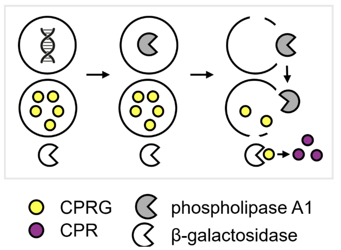
The Colorimetric pH Sensor Implementation combines the pH Cascade, which is made of pH Sensing Cells, substrate liposomes (Substrate LUV: CPRG or Substrate SUV: CPRG) and the LacZ enzyme. It produces a visible color change at pH 6.3 and none at pH 7.6, in solution and in 3.5 mg/mL low-gelling agarose, which is read with Colorimetric Readout. Acidic pH triggers expression of phospholipase A1 (PLA1), which lyses the CPRG liposomes, allowing β-galactosidase to convert released CPRG from yellow to purple. Wells hold 60 µL and are incubated at 37 °C.
Protocol
Solution Assay
The solution assay runs in a 384-well square plate with 60 µL per well. The pH 7.6 arm is plate column 4, and the pH 6.3 arm is plate column 5. The pH 6.3 arm replaces the Tris1M-HEPES1.15M buffer stock with a buffer mix for pH 6.3. The composition of the buffer mix is in the Outer Solution tab of the pH Cascade.
Assembly of a 60 µL well in the solution assay.
| Component | pH 7.6 arm (µL) | pH 6.3 arm (µL) |
|---|---|---|
| Energy solution (2×) | 30 | 30 |
| Ultrapure water | 15.6 | 15.6 |
| Tris1M-HEPES1.15M buffer stock | 7.2 | |
| Buffer mix for pH 6.3 | 7.2 | |
| pH Sensing Cells, CPRG liposomes and suspension buffer | 7.2 | 7.2 |
| Total | 60 | 60 |
The last row of the table above is a 7.2 µL slot that holds the cells, the liposomes and suspension buffer. The table below gives its split for the four wells that were imaged.
Conditions of the four imaged wells. The suspension buffer is 466.7 mL/L Tris1M-HEPES1.15M.
| Well | Name | Type | pH | pH Sensing Cells (µL) | CPRG liposomes (µL) | Suspension buffer (µL) |
|---|---|---|---|---|---|---|
| D4 | pH 7.6, cells only | Control | 7.6 | 1 | 0 | 6.2 |
| D5 | pH 6.3, cells only | Control | 6.3 | 1 | 0 | 6.2 |
| E4 | pH 7.6, cells and liposomes | Sample | 7.6 | 3 | 2.1 | 2.1 |
| E5 | pH 6.3, cells and liposomes | Sample | 6.3 | 3 | 2.1 | 2.1 |
The cells-only wells take 1 µL of cells, and the sample wells take 3 µL, so the two differ in cell dose as well as in liposomes. The four wells are imaged by fluorescence microscopy after 13 h at 37 °C.
Gel Assay
The gel assay uses 60 µL of low-gelling agarose gel per well, set by Embedding: Thermal Setting. It is the solution well with the 15.6 µL of ultrapure water replaced by 15.6 µL of a 27 mg/mL low-gelling agarose stock, which gives a final agarose concentration of 7 mg/mL. The pH 6.3 arm takes the same buffer mix as in the solution assay.
Assembly of a 60 µL well in the gel assay, at 7 mg/mL agarose.
| Component | pH 7.6 arm (µL) | pH 6.3 arm (µL) |
|---|---|---|
| Energy solution (2×) | 30 | 30 |
| 27 mg/mL agarose stock in ultrapure water | 15.6 | 15.6 |
| Tris1M-HEPES1.15M buffer stock | 7.2 | |
| Buffer mix for pH 6.3 | 7.2 | |
| pH Sensing Cells, CPRG liposomes and suspension buffer | 7.2 | 7.2 |
| Total | 60 | 60 |
The agarose stock is 40.5 mg of low-gelling agarose (Sigma-Aldrich A9414-5G) in 1500 µL of ultrapure water. It is dissolved in a heat block at 85 °C to 95 °C with occasional vortexing, then cooled to 65 °C. The stock is mixed with the energy solution and the buffer, the mixture is cooled to 45 °C, and the cells and liposomes are added last. For 3.5 mg/mL agarose, half of the stock is replaced with ultrapure water: 7.8 µL of stock and 7.8 µL of water in place of the 15.6 µL.
The 7.2 µL slot holds 3 µL of pH Sensing Cells, 3 µL of CPRG liposomes and 1.2 µL of suspension buffer. The suspension buffer is at 1175 mOsm/kg and pH 8.
Readout
Color is developed with Colorimetric Readout, which adds 30 µL of a 3× β-galactosidase solution to 60 µL of gel or solution. The 30 µL of 3× solution gives 1.2 U/mL, as in the run in 3.5 mg/mL agarose. The first gel run used 2 U/mL, and its preparation is not given.
Performance
Validation in Solution
In solution, acidic pH gave a visible color change.
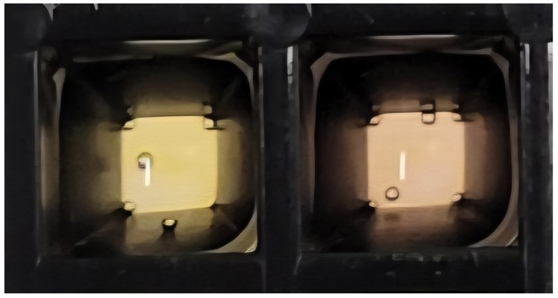
Photograph of the two-liposome system in solution at pH 7.6 (left) and pH 6.3 (right).
Fluorescence microscopy shows the cells in four wells after 13 h at 37 °C. Note: The wells lacking CPRG liposomes (D4 and D5) hold approximately 30% of the number of cells in the sample wells (E4 and E5), so brightness of samples at the same pH are not directly comparable.
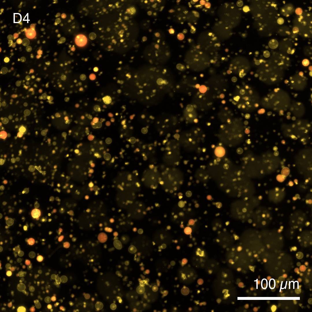
Fluorescence micrograph of pH Sensing Cells at pH 7.6 after 13 h at 37 °C. Cy5 signal stays inside the cells, which indicates that the membranes stayed intact.
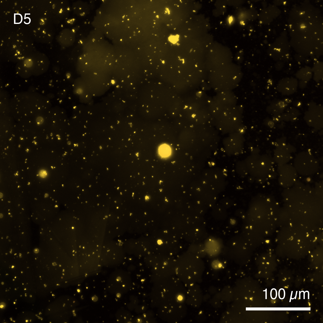
Fluorescence micrograph of pH Sensing Cells at pH 6.3 after 13 h at 37 °C. Cy5 signal is absent, which indicates that acidic conditions triggered PLA1 expression, which disrupted the membrane and released the dye.
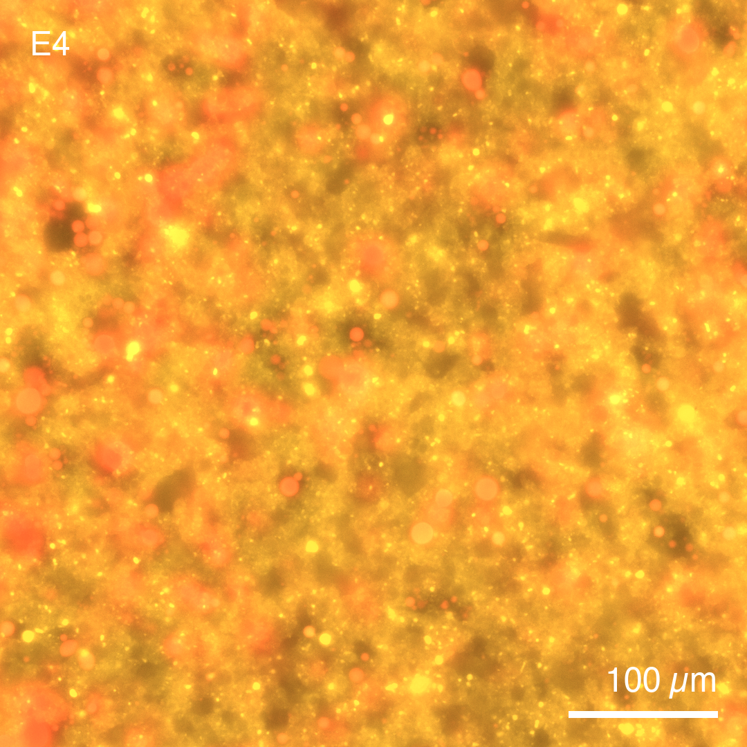
Fluorescence micrograph of CPRG liposomes with pH Sensing Cells at pH 7.6 after 13 h at 37 °C. Cy5 signal is detected only in the cells.
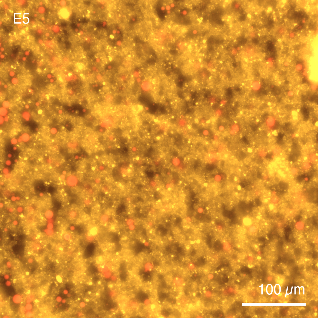
Fluorescence micrograph of CPRG liposomes with pH Sensing Cells at pH 6.3 after 13 h at 37 °C. Cy5 signal is detected only in the cells. In the field shown, fewer cells keep Cy5 than in E4, which indicates PLA1-mediated Cy5 release.
Validation in Gels
CPRG LUVs in 7 mg/mL low-gelling agarose showed a visible color change at pH 6.3, but was barely distinguishable from the negative control lacking pH Sensing Cells, which also developed color. The only difference from the solution assay was the gel, implying that gel embedding caused CPRG to leak from the LUVs, even without PLA1.
To lower the CPRG leakage, the LUVs were extruded through 400 nm membranes to make SUVs, because smaller liposomes are expected to be more stable. Background color change was slightly lower than with LUVs, but the difference between on and off states was still not easily distinguishable by eye.
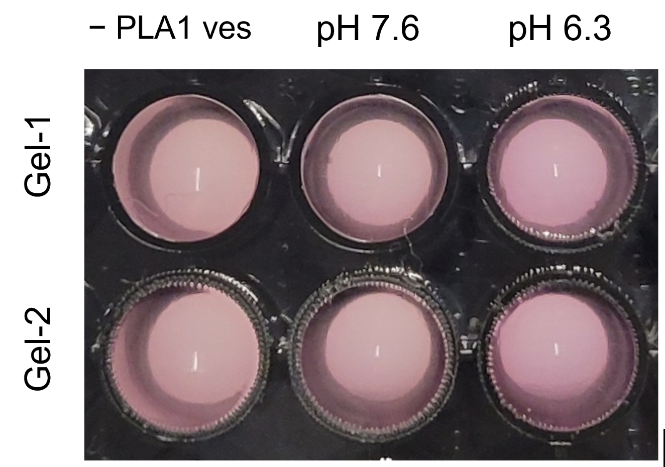
Photograph of color development after 2 h at 37 °C with 2 U/mL β-galactosidase. Two gel replicates (Gel-1 and Gel-2) each show the control without pH Sensing Cells (− PLA1 ves) and both pH arms.
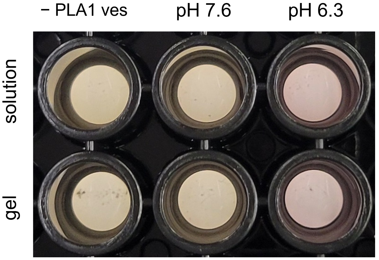
Photograph of color development after 5.5 h at 37 °C with 1.2 U/mL β-galactosidase, in solution and in gel. The control without pH Sensing Cells and the pH 7.6 arm stay yellow in both rows. The pH 6.3 arm turns pink in both rows.
The starting volume of the SUV preparation was raised to increase the yield. More washes were done to lower the background color change. The agarose concentration was lowered from 7 mg/mL to 3.5 mg/mL to reduce liposome lysis during gel embedding. A solution assay was run in parallel as a control. Color change was visible in both gel and in solution.
Process
Anneal pH Trigger Duplex makes the gate duplex of the pH Sensing Cell.
Encapsulation: Freeze-Thaw makes the CPRG liposomes as LUVs, and as SUVs from the second gel run onward.
Protocol: Inverted Emulsion makes the pH Sensing Cells.
Embedding: Thermal Setting places the cells and liposomes in a gel.
Colorimetric Readout develops and reads the color.
Modules
Credits
Developed by Sung-Won Hwang, Samuel Chen and Allen P. Liu (University of Michigan).
Supported by
DevNotes that support this page include: