Overview
The Base Cell is Base Cytosol encapsulated in Base Membrane via Encapsulation: Phase Transfer. The Base Cell is deployed in a glucose outer solution and represents the default synthetic cell for the Nucleus distribution. Base Cells have red fluorescent membranes (Liss-Rhod PE) and express GFP over time if functioning correctly.
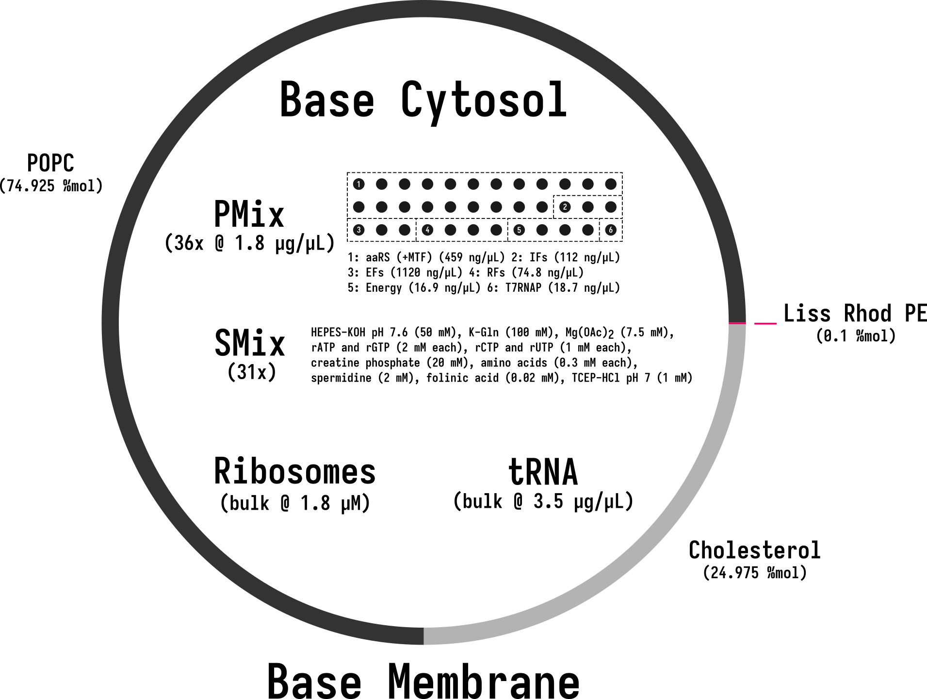
Overview of Base Cell, composed of Base Cytosol encapsulated with Base Membrane. Constitutive components of each module schematically depicted.
Reference Composition
The inner solution encapsulated into the Base Cell is Base Cytosol at reaction concentration, with pOpen-deGFP DNA added as a reporter.
| Component | Input concentration | Final concentration | Volume for one reaction (µL) |
|---|---|---|---|
| SMix | 3.33x | 1x | 12 |
| PMix | 15 mg/mL | 1.80 mg/mL | 4.8 |
| Ribosomes | 10 µM | 1.8 µM | 7.2 |
pOpen-deGFP DNA | 124 nM | 3 nM | 0.95 |
| tRNA | 35 mg/mL | 3.5 mg/ml | 4 |
| Optiprep | 1.32 mg/µL | 0.043 mg/µL | 1.33 |
| RNase Inhibitor | 40 000 U/mL | 2000 U/mL | 2 |
| Water | 6.12 | ||
| Total volume (µL) | 40 |
| Component | Target Percentage (%) | Molecular Weight (g/mol) | Stock concentration (mg/mL) | Volume to add (µL) |
|---|---|---|---|---|
| POPC | 70 | 760.076 | 25 | 162.17 |
| Cholesterol | 29.95 | 386.654 | 50 | 17.65 |
| Liss-Rhod PE | 0.05 | 1301.71 | 1 | 4.96 |
See Base Membrane for the full membrane spec.
| Component | Concentration |
|---|---|
| Glucose | 850 mM |
Expected Behavior
Base Cells express deGFP over time, starting dark and increasing in green fluorescence as translation proceeds. Kinetics data are reported in DevNote bnext-devnotes-base-cell-01.
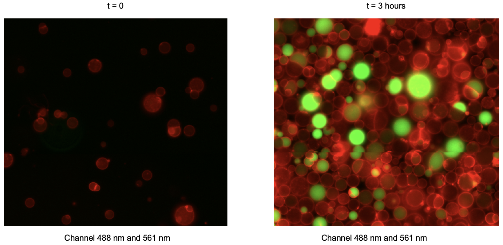
Combined green (488 nm) and red (561 nm) fluorescence channels. First time point (labeled “t = 0”) corresponds to 30 min after preparation of the inner solution, reflecting the time required to prepare liposomes.
Process
Base Cells are assembled and encapsulated using Encapsulation: Phase Transfer.
Constituent Modules
Base Cytosol — inner solution, encapsulated at reaction concentration
Base Membrane — 70:30 POPC:cholesterol bilayer
Credits
Developed by Surendra Yadav (b.next).