Overview
The Cx43 Module expresses connexin 43 (Cx43), a mammalian gap junction protein, that self-assembles into hexameric hemichannels (connexons) that spontaneously integrate into the membrane of synthetic cells and permit the passage of molecules up to ~1 kDa. Cx43 provides an alternative to α-hemolysin: its status as a non-select agent makes it easier to distribute, and its capacity to form gap junctions between neighboring cells represents a new function that opens a path toward tissue-like assemblies.
This Module was contributed to the Nucleus Community by Ahmed Sihorwala (Belardi Lab, UT Austin), based on the construct design from the Stachowiak Lab and the publication Sihorwala et al., 2023. Validation data is presented in the DevNote Cx43 Cell: DNA Validation.

Depiction of connexin and its relationship to a connexon: (left) Connexins are membrane-spanning proteins whose N- and C-termini are located in the cytoplasm; (right) connexons are complexes of six connexins. Figure by Totland et al., 2023 used under CC-BY-4.0 / cropped from original.
| Construct | Size | Description | File |
|---|---|---|---|
pOpen-pT7-Cx43 | 3320 bp | Expresses wild-type Cx43 under T7 promoter in pOpen backbone | pOpen-Cx43.gb |
pOpen-pT7-Cx43-eGFP | 3996 bp | Expresses Cx43-eGFP fusion protein under T7 promoter in pOpen backbone | pOpen-Cx43-eGFP |
Cytosol
Cx43 requires a lipid membrane environment for proper folding and pore assembly. To the best of our knowledge, this Module cannot be functionally deployed in Nucleus Cytosol alone.
Cells
Expected Performance
Insertion Assay — Liposomes encapsulating NEB PURExpress and pOpen-pT7-Cx43-eGFP were incubated at 37°C for 6 hrs. Green fluorescent rings around liposomes confirm membrane localization of Cx43-eGFP. Control liposomes lacking the Cx43 plasmid show no rings.
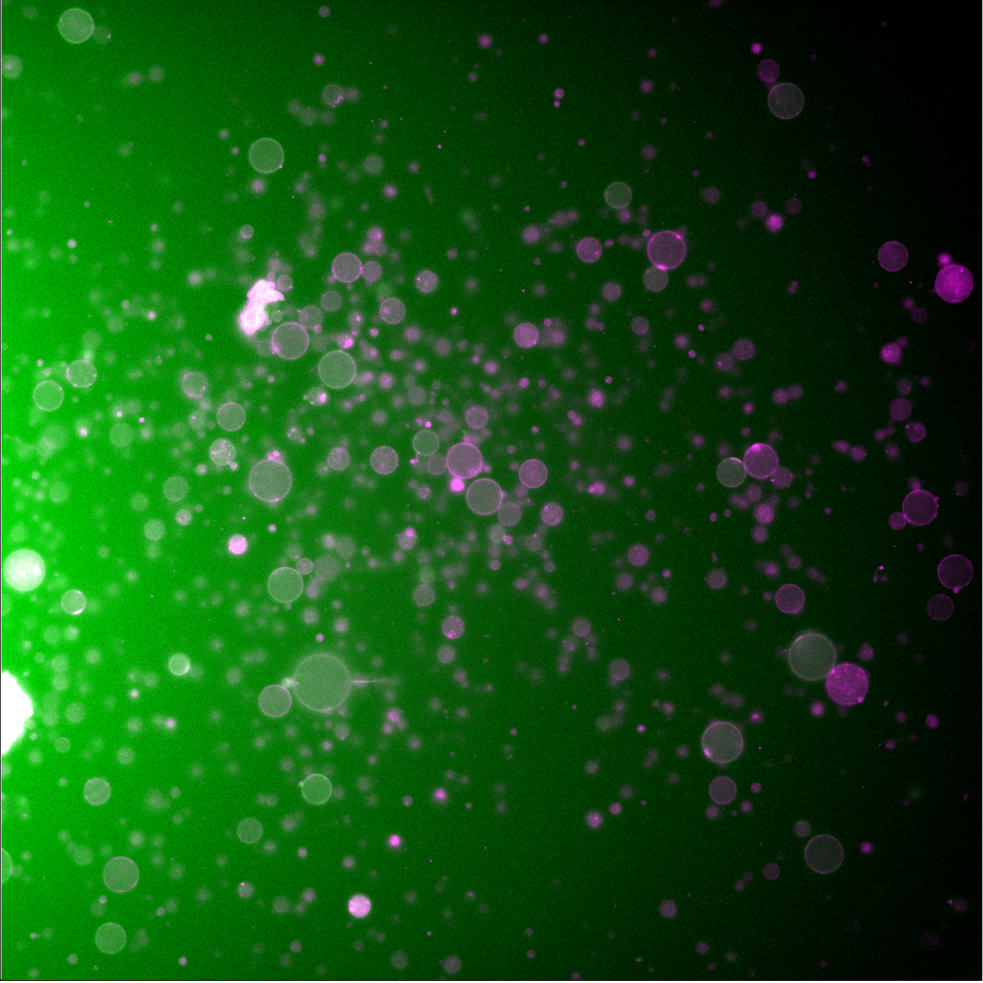
Liposomes expressing Cx43-eGFP. Green fluorescent rings surrounding liposomes indicate successful membrane localization of Cx43-eGFP hemichannels. 488 nm (green, Cx43-eGFP) and 561 nm (red, membrane label) channels overlaid.
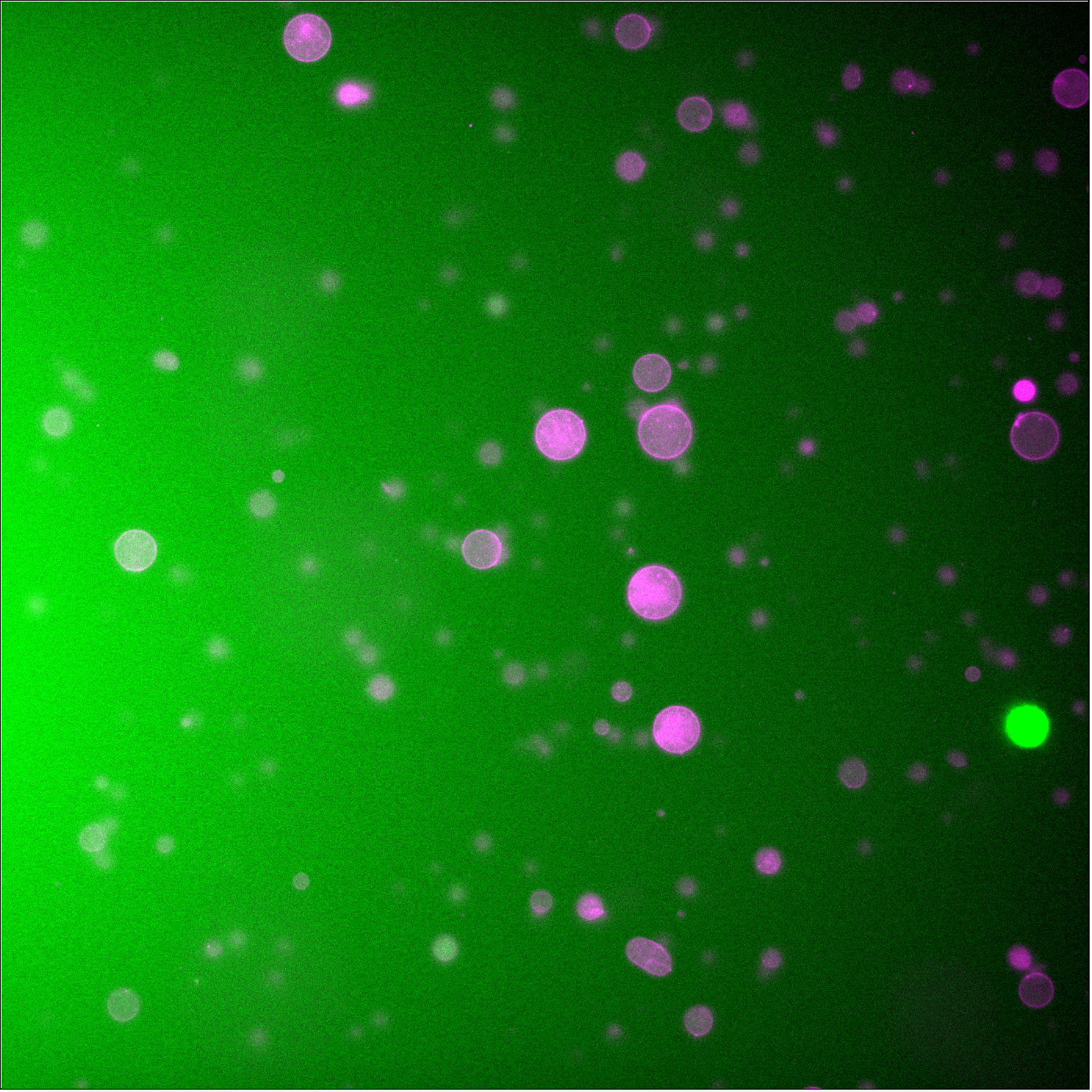
Control liposomes without Cx43 plasmid. No green fluorescent rings are observed. Faint green signal within liposomes is encapsulated PURE.
Leakage Assay — Liposomes co-encapsulating NEB PURExpress, pOpen-pT7-Cx43, and Alexa Fluor 647 dye were incubated at 37°C for 6 hrs and imaged by confocal microscopy every 10 min.
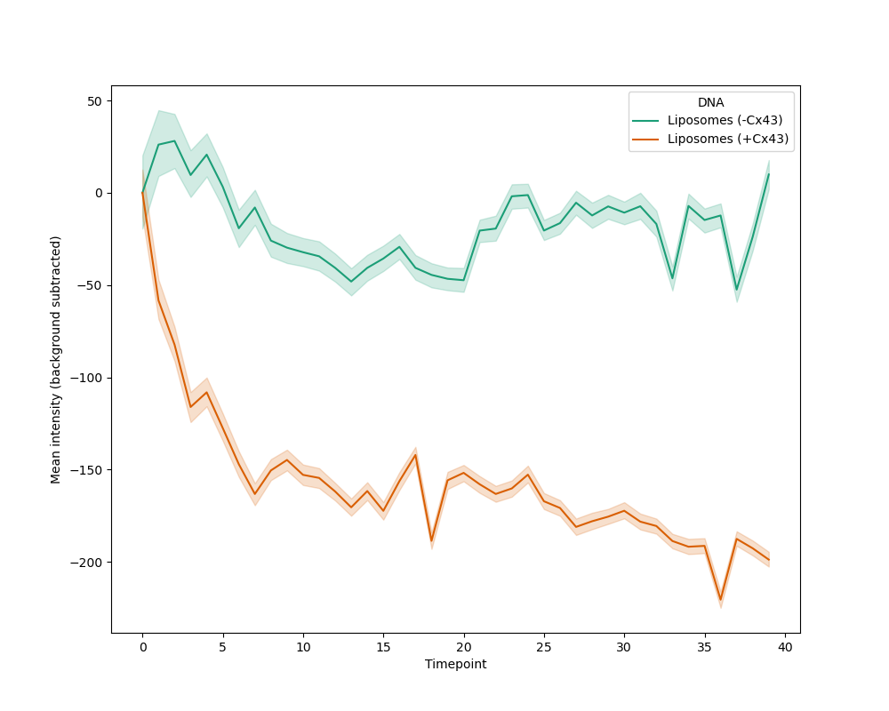
Background-subtracted Alexa Fluor 647 fluorescence intensity over 6 hrs at 37°C. Liposomes containing Cx43 show a progressive decrease in encapsulated dye fluorescence relative to controls, consistent with pore-mediated dye leakage.
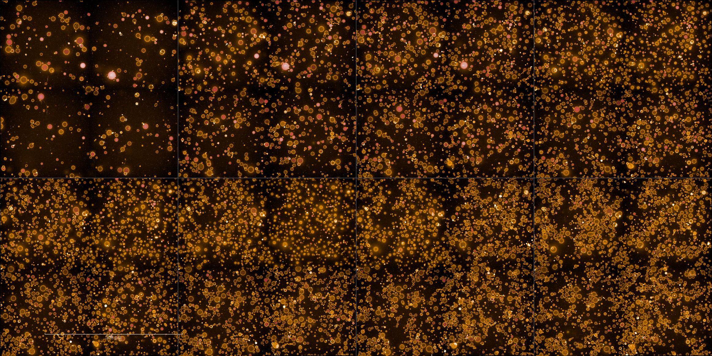
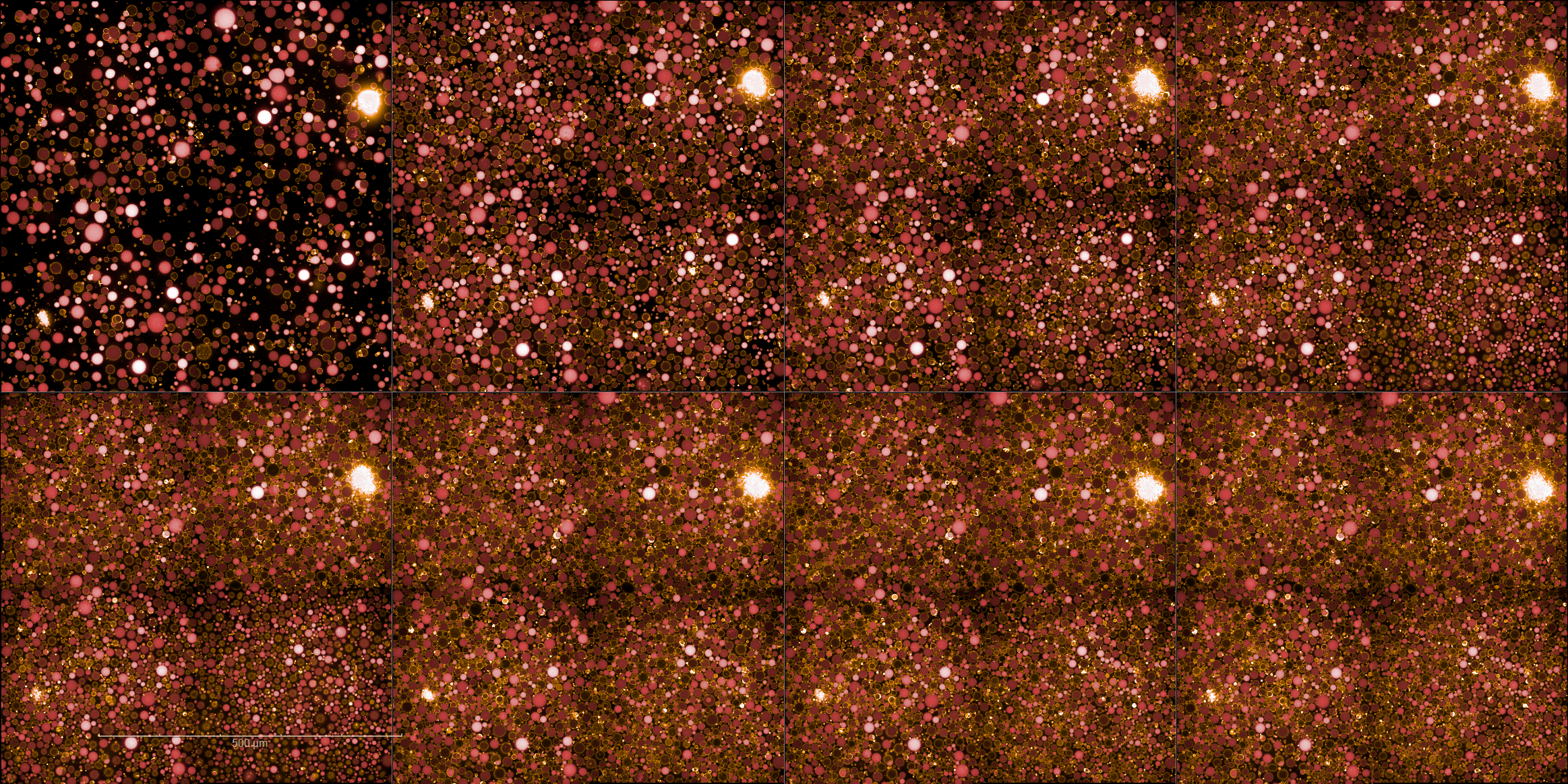
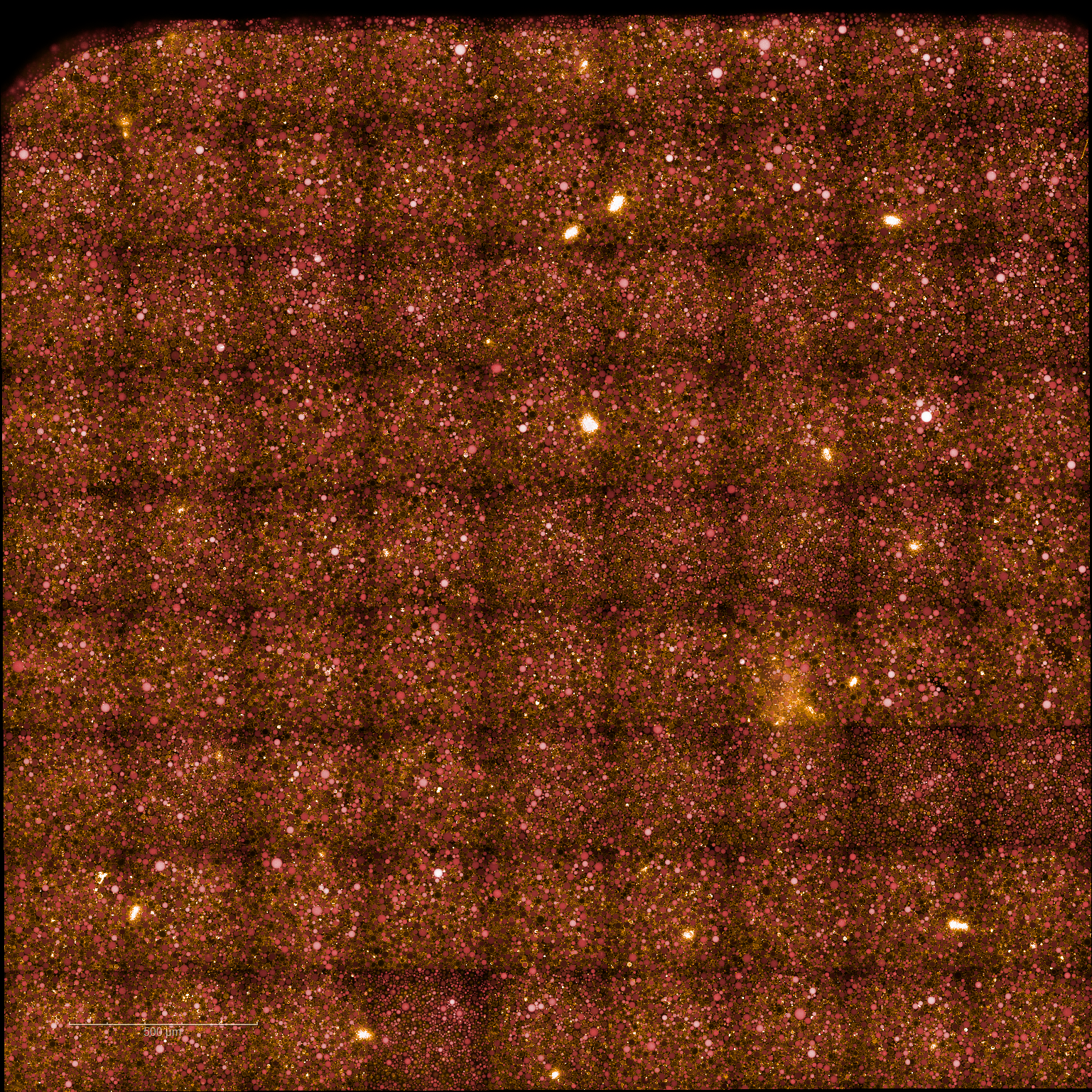
Time series of Cx43-reconstituted liposomes encapsulating Alexa Fluor 647 over 6 hrs at 37°C (images every 10 min, starting 40 min after preparation). Progressive loss of fluorescence is observed as dye leaks through Cx43 channels. Scale bar: 500 µm.
Time series of control liposomes encapsulating Alexa Fluor 647 over 6 hrs at 37°C. Dye fluorescence is maintained throughout, confirming that leakage requires Cx43 expression. Scale bar: 500 µm.
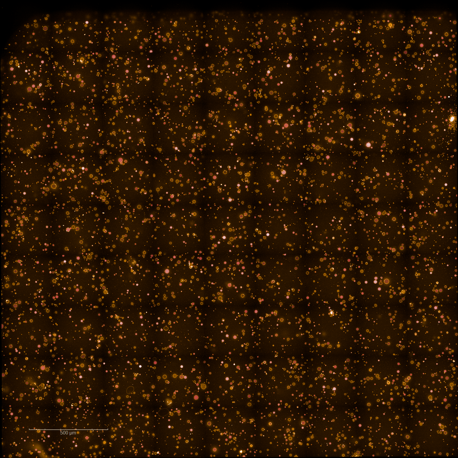
Confocal image of Cx43-reconstituted liposomes at the start point, 40 min after preparation. Some liposomes already show reduced fluorescence, consistent with early dye leakage at room temperature. Scale bar: 500 µm.
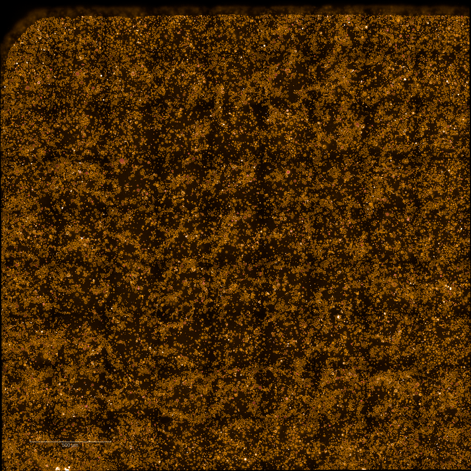
Endpoint confocal image (6 hrs 40 min) of Cx43-reconstituted liposomes. A higher proportion of non-fluorescent liposomes is observed relative to controls. Scale bar: 500 µm.
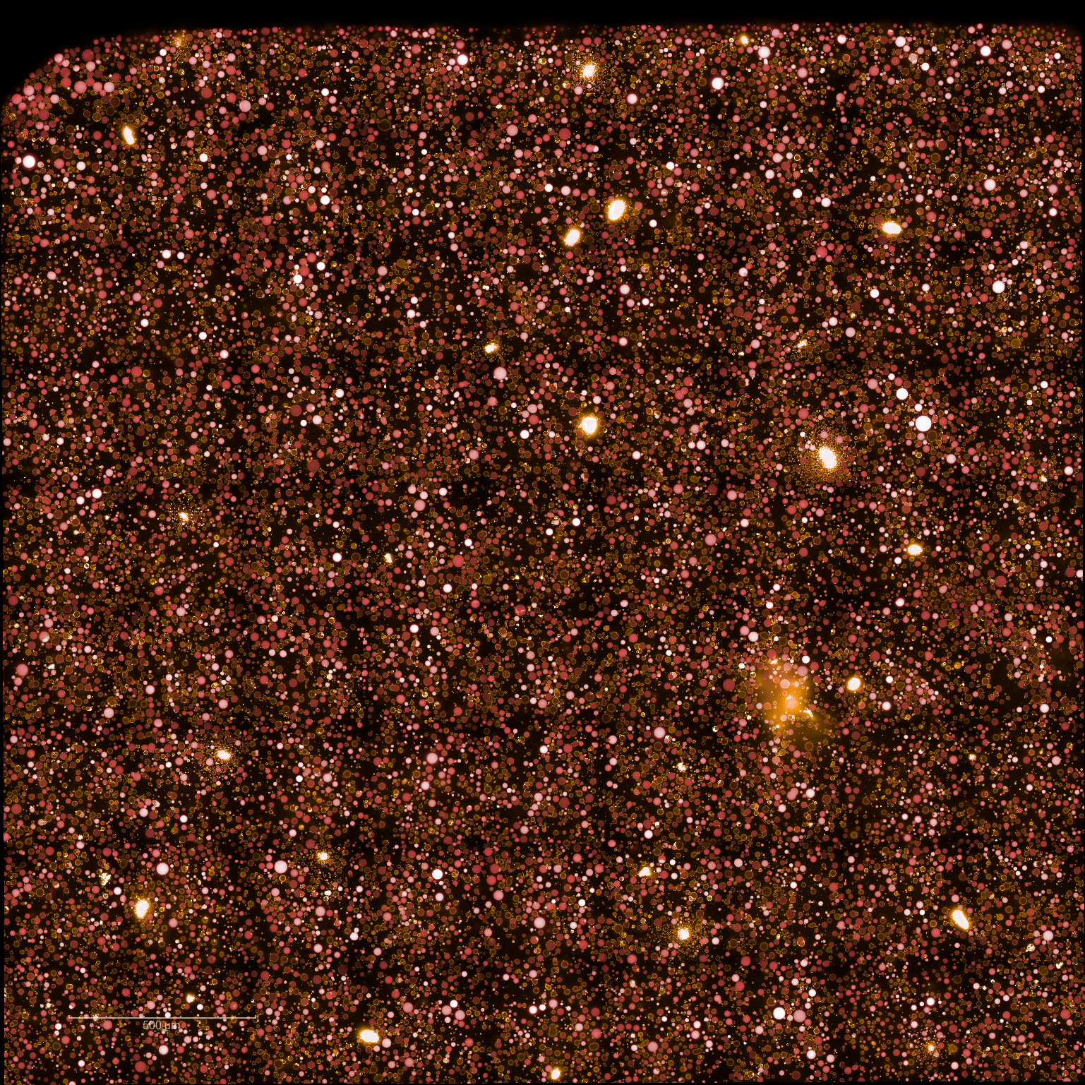
Confocal image of control liposomes at the start point, 40 min after preparation. Liposomes remain fluorescent, confirming dye retention in the absence of Cx43. Scale bar: 500 µm.
Endpoint confocal image (6 hrs 40 min) of control liposomes. Most liposomes remain fluorescent throughout the experiment. Scale bar: 500 µm.
Credits
Module contributed by Ahmed Sihorwala (Belardi Lab, UT Austin). Validation data by Yen-Yu Hsu (b.next).
- Sihorwala, A. Z., Lin, A. J., Stachowiak, J. C., & Belardi, B. (2023). Light-Activated Assembly of Connexon Nanopores in Synthetic Cells. Journal of the American Chemical Society, 145(6), 3561–3568. 10.1021/jacs.2c12491
- Hsu, Y.-Y. (2025). Cx43 Cell: DNA Validation. 10.63765/xvxu3274
- Totland, M. Z., Omori, Y., Sørensen, V., Kryeziu, K., Aasen, T., Brech, A., & Leithe, E. (2023). Endocytic trafficking of connexins in cancer pathogenesis. Biochimica et Biophysica Acta (BBA) - Molecular Basis of Disease, 1869(7), 166812. 10.1016/j.bbadis.2023.166812