Overview
The TetR inducible expression module is a set of two genetic constructs that encode tetracycline-inducible gene expression: pT7-tetR, encoding the TetR repressor protein, and pT7-tetO-plamGFP, encoding a reporter gene under an inducible T7 promoter.
pT7-tetO-plamGFP constitutively expresses the open reporter plamGFP in the absence of repressor protein. The inducible promoter is also a MoClo Level 0 ‘P’ part and may be assembled into a Level 1 transcription unit with other MoClo-compatible genes. Addition of TetR protein — either as a purified protein or via constitutive expression of pT7-tetR — inhibits expression through steric occlusion of the tetO operator site. Addition of anhydrotetracycline (aTc) causes allosteric release of TetR from tetO, recovering expression. aTc is membrane-permeable, so the alpha-hemolysin membrane pore is not required for induction.
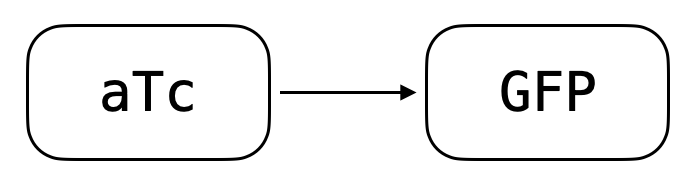
Schematic of the TetR inducible expression module. TetR represses expression from pT7-tetO-plamGFP; aTc relieves repression by binding TetR and causing its release from the tetO operator.
Reference Composition
| Name | Length (bp) | File |
|---|---|---|
pT7-tetR | 2877 | pOpen-tetR.gb |
pT7-tetO-plamGFP | 2954 | pOpen-pT7-tetO.gb |
Assemble pT7-tetO-plamGFP into a standard PURE reaction. Add purified TetR protein to a final concentration of 500 nM, or include the pT7-tetR DNA construct. Add aTc inducer at 2.5 µM to 5 µM for effective induction. Volumes in µL.
| Component | Master Mix (µL) |
|---|---|
| PURExpress Solution A | 4 |
| PURExpress Solution B | 3 |
| RNase Inhibitor | 0.5 |
pT7-tetO-plamGFP (10 nM) | 0.5 |
| TetR (10 µM) | 0.5 |
| Master Mix Total | 9 |
| Component | Per Reaction (µL) |
|---|---|
| Master Mix | 9 |
| Inducer | 1 |
| Total | 10 |
Expected Behavior
Cytosols
The TetR module was validated in NEB PURExpress reactions. Purified repressor protein (MedChemExpress, HY-P71520A) and anhydrotetracycline inducer (Cayman Chemical, 10009542) were added at the final concentrations indicated. pT7-tetO-plamGFP plasmid DNA was added at 0.5 nM.
Repression follows a roughly linear trend between 125 and 750 nM TetR and saturates around 500 nM, though it can be further improved up to 2000 nM. An inducer concentration of 2.5 µM to 5 µM provides effective induction well below saturating or toxic aTc levels. Note that aTc’s yellow color overwhelms GFP fluorescence at concentrations greater than 50 µM to 100 µM, and high concentrations may negatively affect expression generally.
In vitro repression with TetR
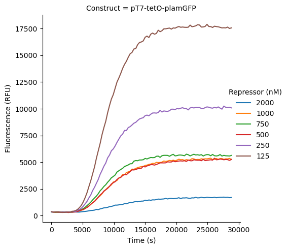
Repression kinetics of pT7-tetO-plamGFP by TetR at varying repressor concentrations.
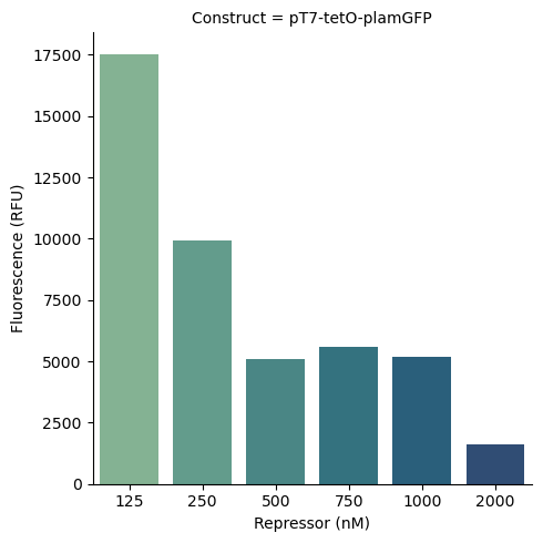
Repression of pT7-tetO-plamGFP by TetR at steady state.
In vitro induction with aTc
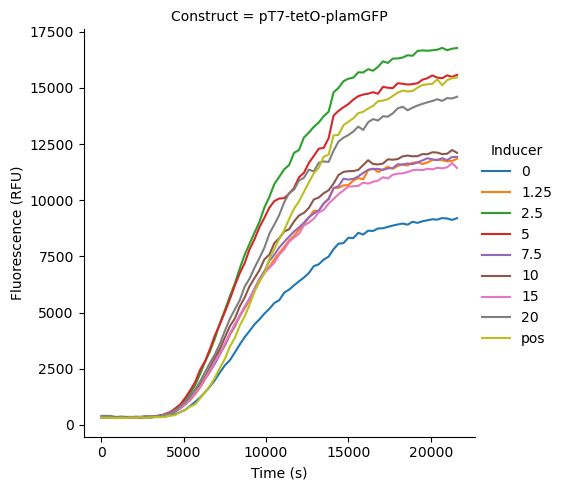
Induction kinetics of pT7-tetO-plamGFP by aTc. TetR repressor protein is present at 500 nM. Positive control is pT7-tetO-plamGFP without TetR repressor protein.
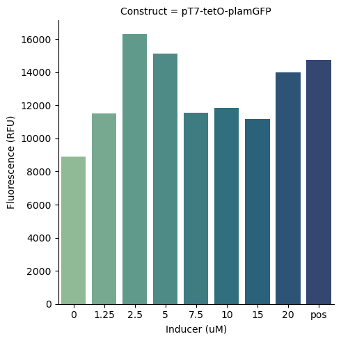
Induction of pT7-tetO-plamGFP by aTc at steady state. TetR repressor protein is present at 500 nM. Positive control is pT7-tetO-plamGFP without TetR repressor protein.
Cells
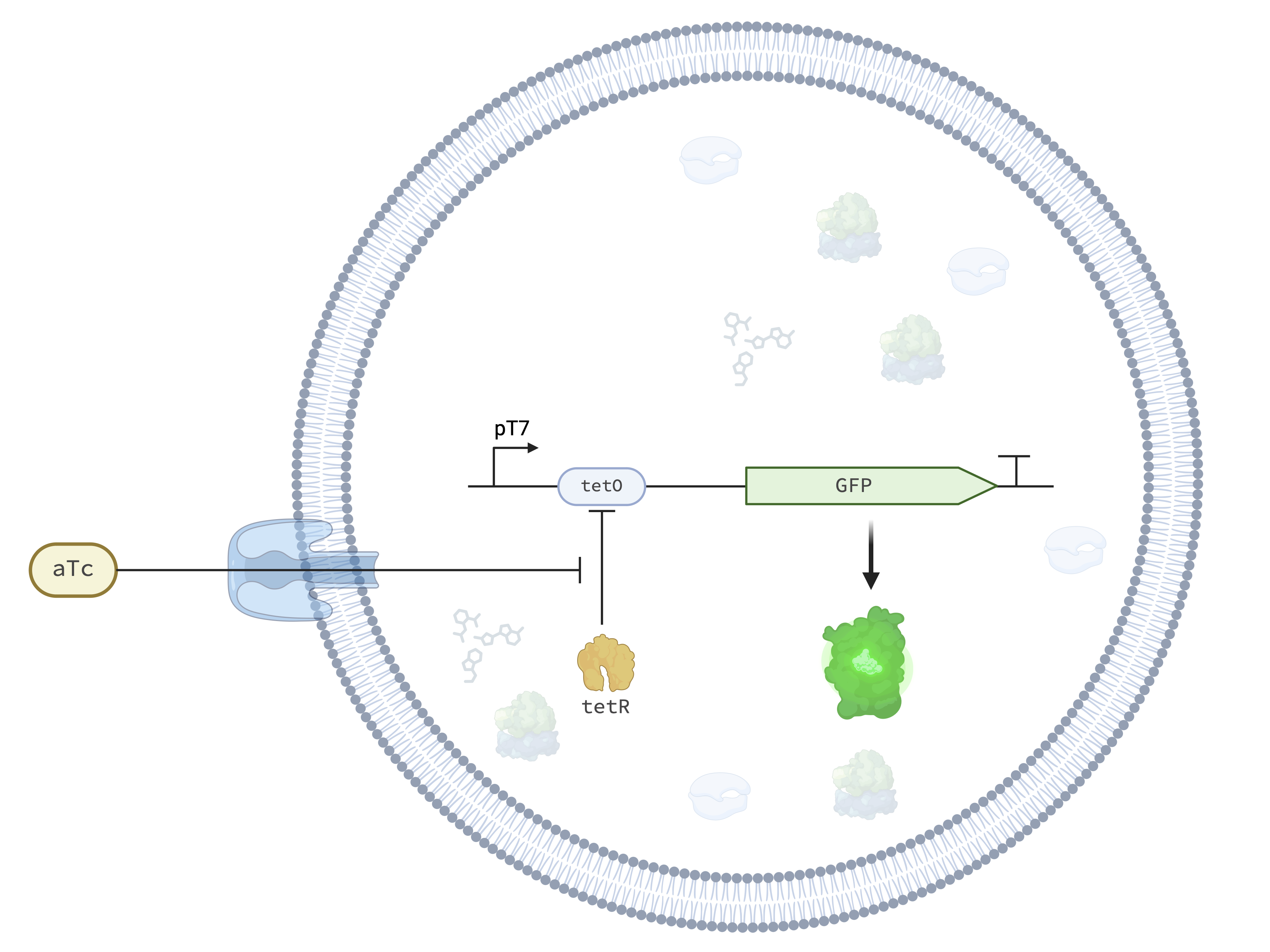
The TetR-aTc Detector module in the Base Cell.
TetR detector synthetic cells were induced at multiple anhydrotetracycline concentrations and imaged over 12 h with approximately 22 min per timepoint.
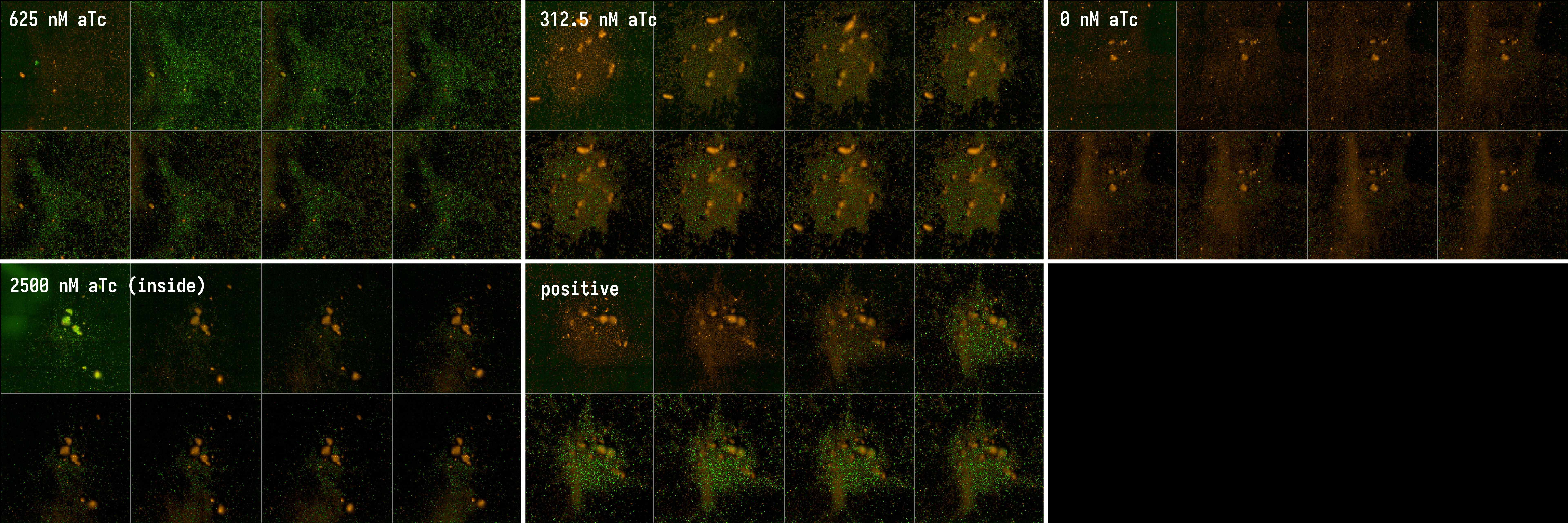
TetR detector synthetic cells induced at multiple anhydrotetracycline concentrations. 8 timepoints displayed per condition, approximately 22 min apart, over 12 h total. First row: induction using 625 nM, 312.5 nM, and 0 nM (fully repressed) aTc introduced into the outer buffer. Second row: induction with 2500 nM aTc in the inner solution and positive control without TetR repression.
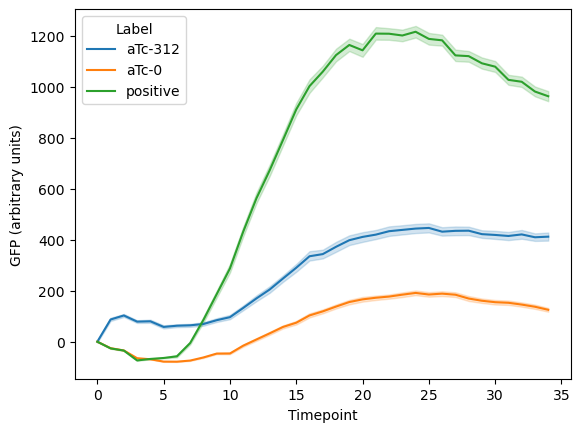
GFP expression within synthetic cells when induced with 312.5 nM anhydrotetracycline.
The TetR detector cell functions when induced with low-nanomolar aTc concentrations. Higher concentrations begin to inhibit expression or confound analysis due to background aTc fluorescence and membrane localization.
Requirements
Requires pT7 transcription and translation (e.g. Base Cytosol).
Implementations
Responder: aTc → IV-HSL: aTc relieves TetR repression to drive BjaI expression.
Credits
Developed by Yen-Yu Hsu (b.next).