Overview
The ClpXP control module enables the ATP-dependent, targeted degradation of ssrA-tagged proteins McGinness, Baker, and Sauer, 2006. It is based on the complex formed by the AAA+ ATPase ClpX and the tetradecameric peptidase ClpP. The module can be implemented using purified protein, in situ expressed proteins from DNA templates, or combinations thereof.
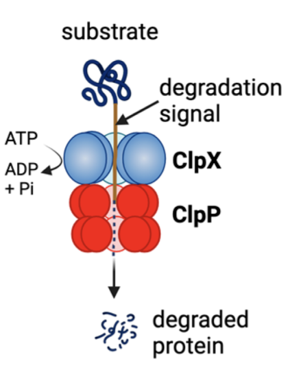
Cartoon of the general mechanism of protein degradation by ClpXP, an ATP-dependent protease. Adapted from R. Wedam, et al.
Reference Composition
The module can be implemented from purified proteins alone, from in situ expressed proteins encoded on DNA templates, or from combinations thereof.
| Name | Length (bp) | File |
|---|---|---|
pT7-ClpX | 3394 | pOpen-ClpX-CHis.gb |
pT7-ClpP | 2746 | pOpen-ClpP-CHis.gb |
pT7-deGFP-ssrA | 2863 | pOpen |
Reaction Table 1. The control module implemented from purified proteins. Volumes in µL.
| Component | Sample 1 | Sample 2 | Sample 3 | Control |
|---|---|---|---|---|
| Purified deGFP-ssrA (41.2 µM) | 0.5 | 0.5 | 0.5 | 0.5 |
| Purified ClpP (79.9 µM) | 0.5 | 0.5 | 0 | 0 |
| Purified ClpX (53.7 µM) | 0.5 | 0 | 0.5 | 0 |
| NEB PURExpress Solution A | 4 | 4 | 4 | 4 |
| NEB PURExpress Solution B | 3 | 3 | 3 | 3 |
| RNase Inhibitor | 0.5 | 0.5 | 0.5 | 0.5 |
| Nucleus Free Water | 1 | 1.5 | 1.5 | 2 |
| Total | 10 | 10 | 10 | 10 |
Reaction Table 2. The control module implemented from in situ expressed proteins. Steady-state levels can be tuned by varying the concentration of in situ expressed ClpXP proteins. Volumes in µL.
| Component | Sample 1 | Sample 2 | Sample 3 | Sample 4 |
|---|---|---|---|---|
| pT7-deGFP-ssrA (63.5 ng/µL) | 0.5 | 0.5 | 0.5 | 0.5 |
| pT7-ClpP (17.5 ng/µL) | 0.4 | 0.4 | 0.6 | 0.8 |
| pT7-ClpX (17.5 ng/µL) | 0.4 | 0.4 | 0.6 | 0.8 |
| NEB PURExpress Solution A | 4 | 4 | 4 | 4 |
| NEB PURExpress Solution B | 3 | 3 | 3 | 3 |
| RNase Inhibitor | 0.5 | 0.5 | 0.5 | 0.5 |
| Nucleus Free Water | 1.6 | 1.2 | 0.8 | 0.4 |
| Total | 10 | 10 | 10 | 10 |
Expected Behavior
Cytosols
Module performance in PURE is documented in the DevNote ClpXP Module Validation in PURE.
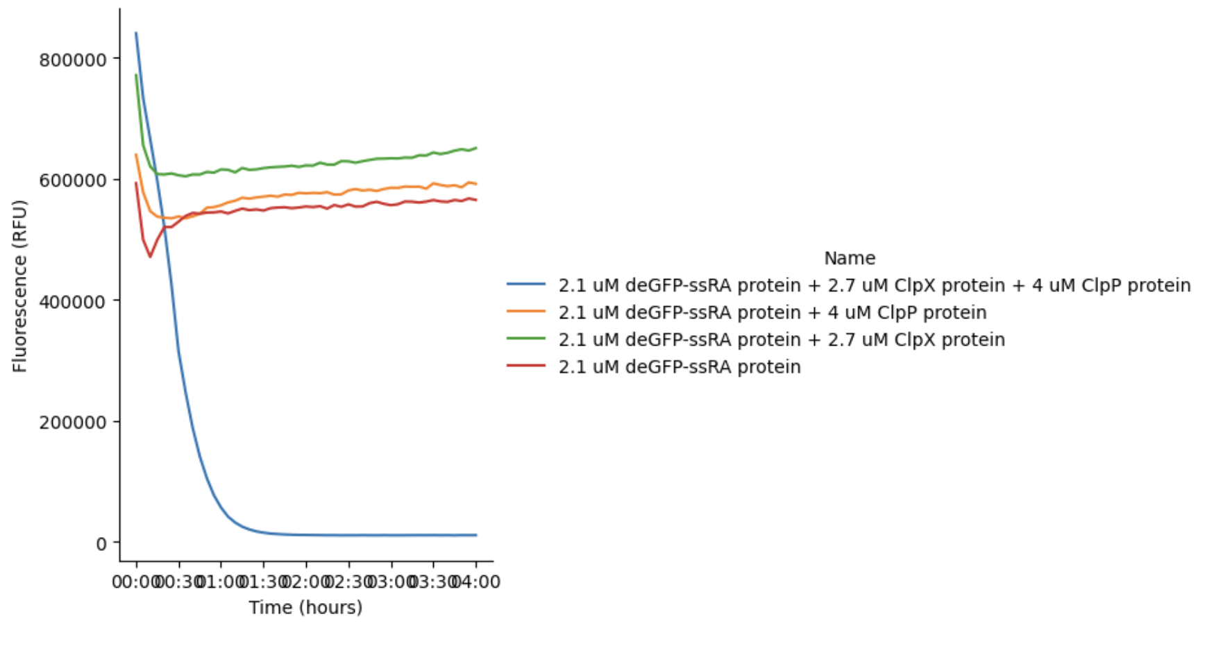
GFP fluorescence of samples containing purified proteins incubated at 37 °C for 4 h. These results correspond to Reaction Table 1.
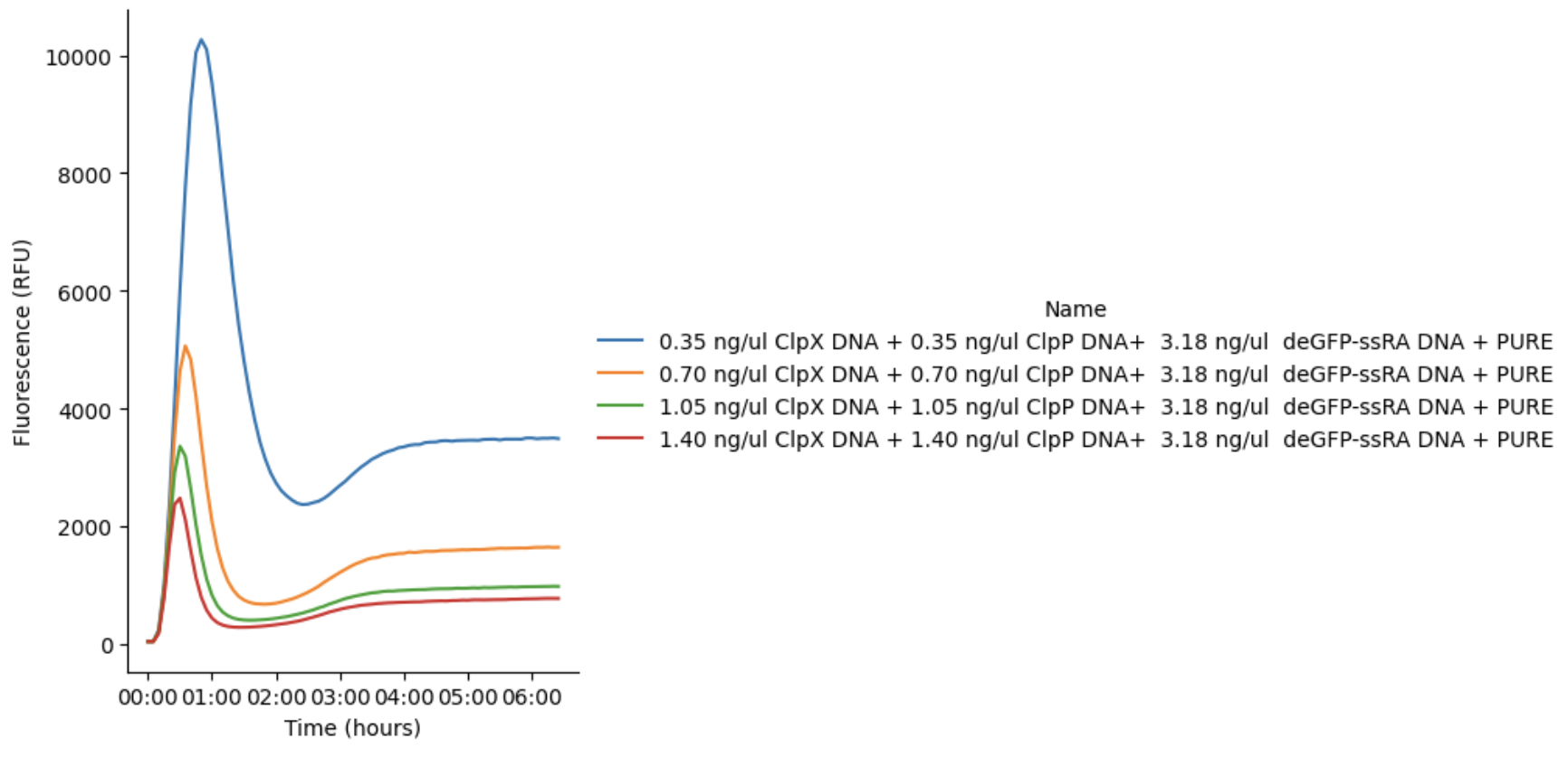
GFP fluorescence signal produced using pT7-deGFP-ssrA DNA in PURE reactions incubated at 37 °C for 6 h. ClpX and ClpP DNAs are co-expressed in the same PURE reaction. These results correspond to Reaction Table 2.
Cells
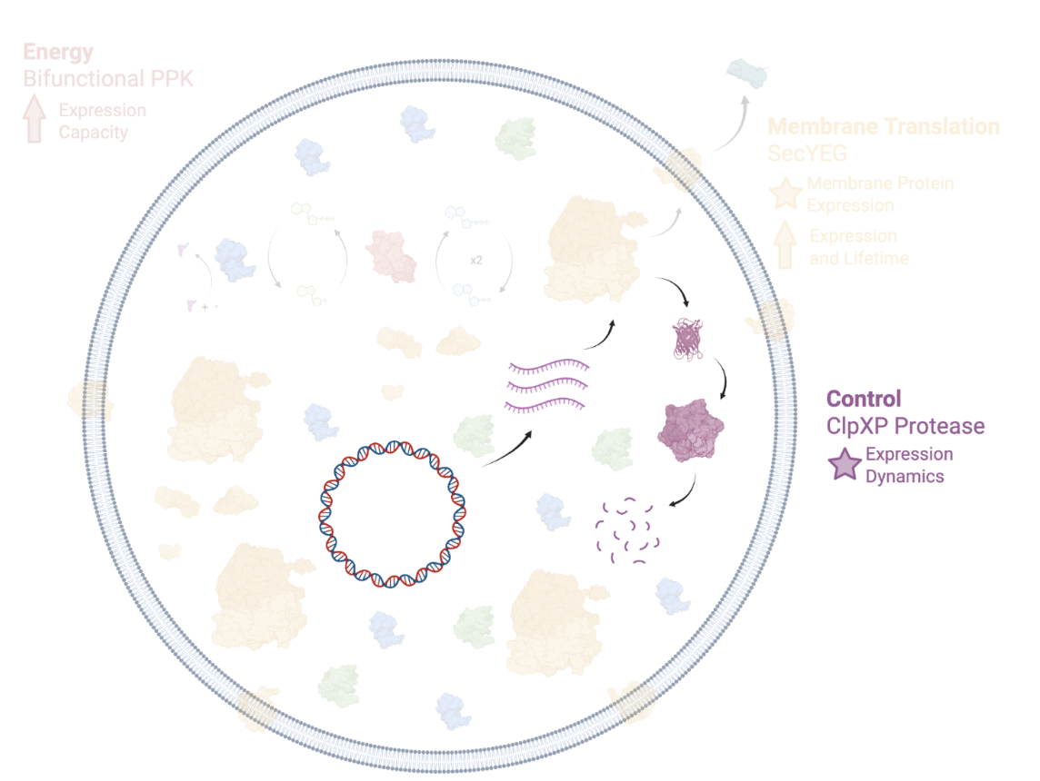
The ClpXP Control Module in the context of the Developer Cell. Other Developer Cell Modules are grayed out.
Cell-context validation of the ClpXP module is documented in the DevNote ClpXP Module Validation in Cells.
Three liposome populations were prepared to test whether the control module functions inside a synthetic cell. All three encapsulated purified deGFP-ssrA together with the PURE system. The first also encapsulated two linear DNAs, pT7-ClpX and pT7-ClpP; the second encapsulated pT7-ClpP DNA with purified ClpX protein; the control contained no DNA.
Liposomes containing functional ClpXP — whether assembled from two co-encapsulated DNAs or from DNA plus purified protein — show a clear decrease in green fluorescence over the incubation. Liposomes carrying both pT7-ClpX and pT7-ClpP degrade GFP more slowly than those carrying only pT7-ClpP with purified ClpX, which is consistent with competition for limited transcription and translation resources when several DNAs share one PURE reaction. Control liposomes show no substantial decrease; the slight reduction that does appear is most likely photobleaching. Expect a modest rise in green fluorescence over the first ~20 min, which reflects liposomes settling to the bottom of the imaging well rather than a change in expression.
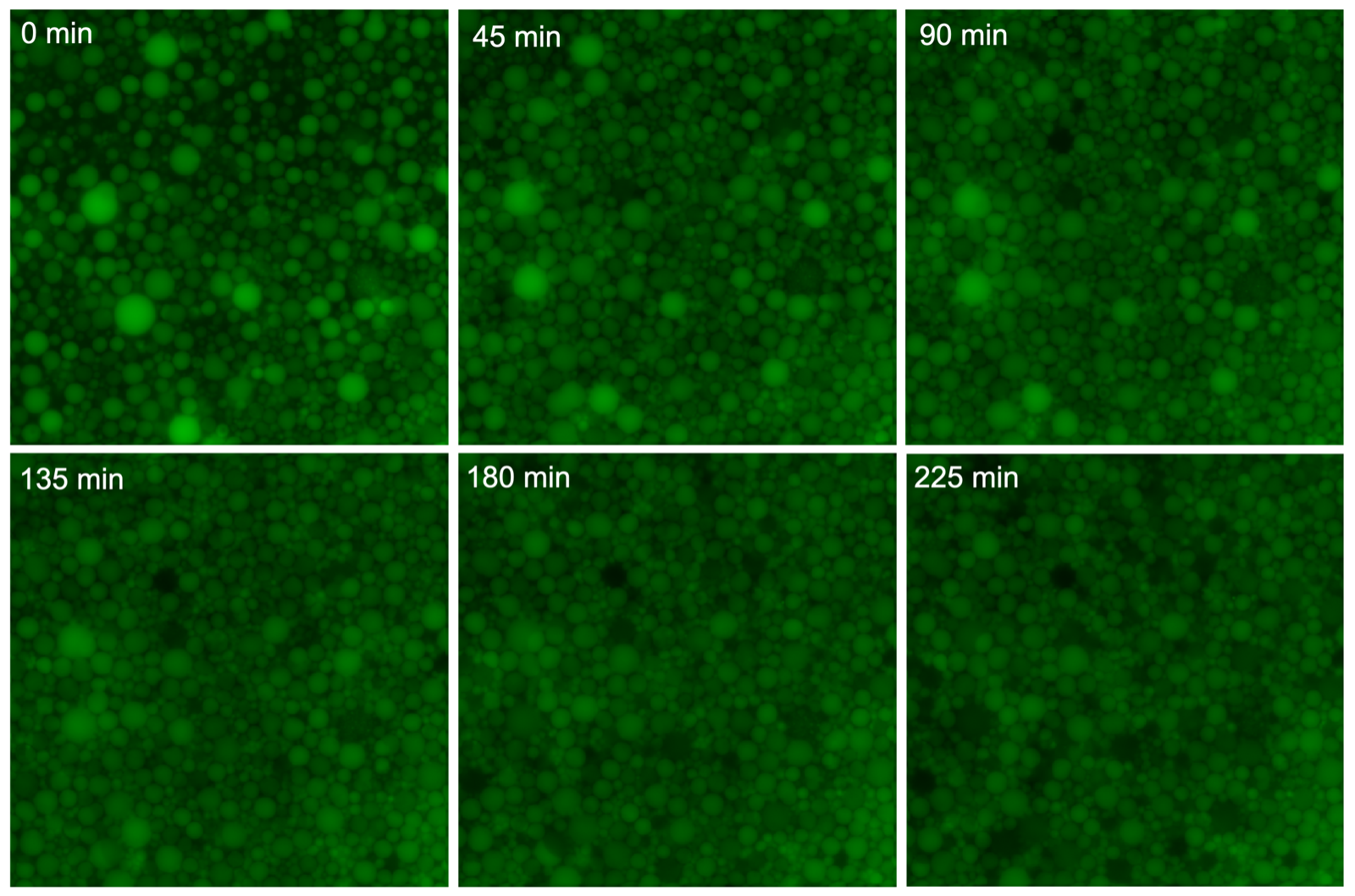
Time-series fluorescence microscopy of liposomes encapsulating pT7-ClpX and pT7-ClpP DNA with purified deGFP-ssrA, incubated at 37 °C. Green fluorescence decreases over time.
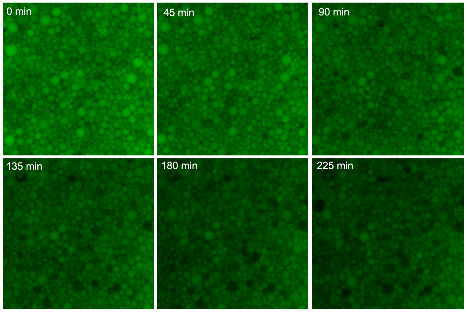
Time-series fluorescence microscopy of liposomes encapsulating pT7-ClpP DNA with purified ClpX and purified deGFP-ssrA, incubated at 37 °C. Green fluorescence decreases faster than in the two-DNA condition.
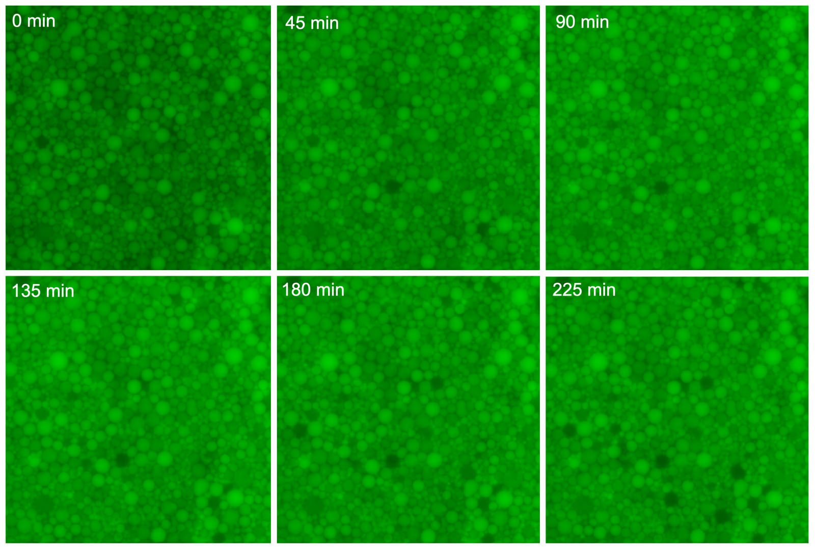
Time-series fluorescence microscopy of control liposomes encapsulating purified deGFP-ssrA only, incubated at 37 °C. Fluorescence stays substantially stable.
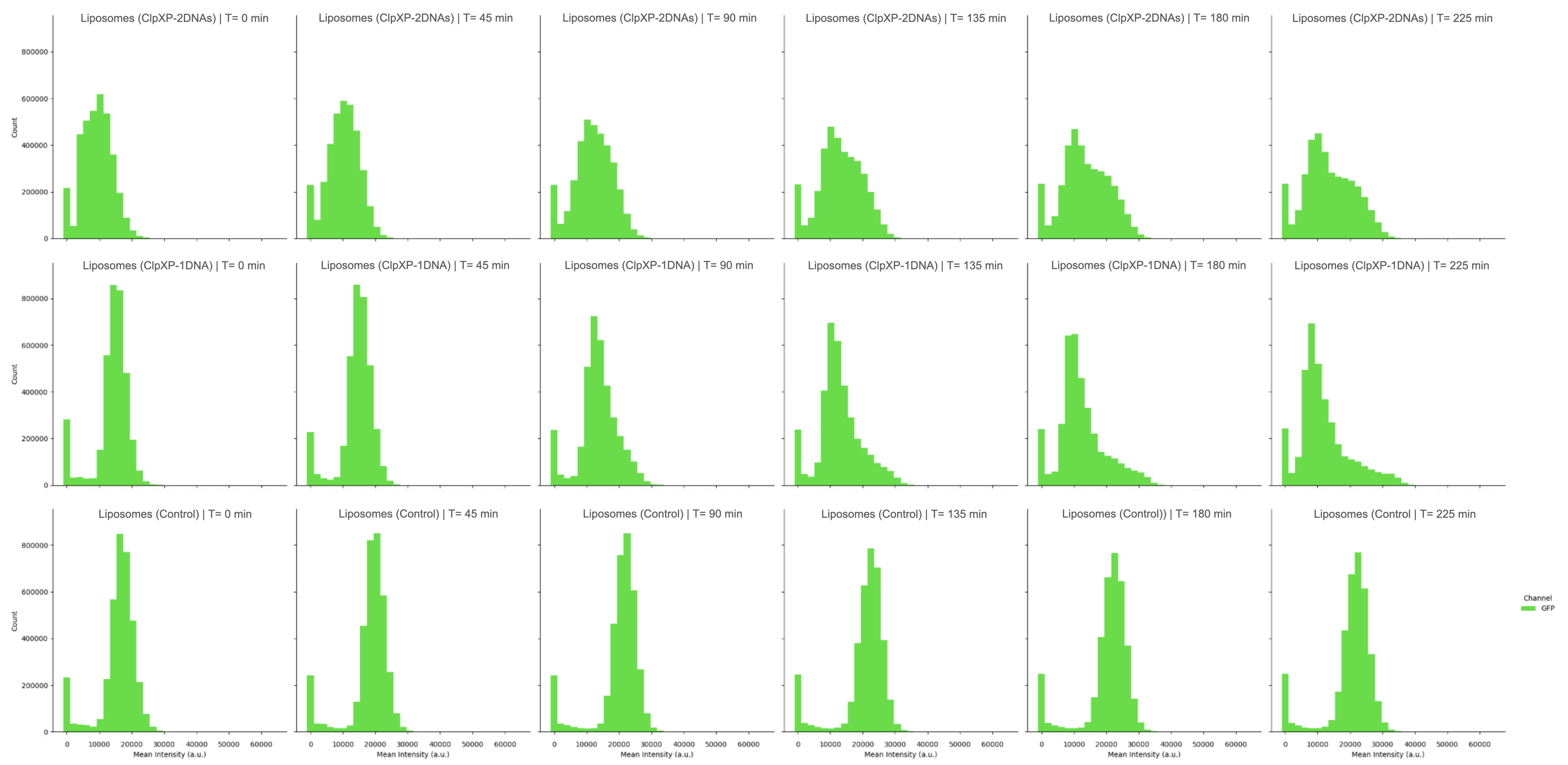
Time-resolved histograms of mean GFP fluorescence intensity for individual liposomes, at 0, 45, 90, 135, 180, and 225 min. Top row: both ClpX and ClpP DNA. Middle row: a single ClpXP DNA component. Bottom row: control liposomes lacking ClpXP. A persistent high-intensity subpopulation appears in all three conditions.
Quantifying single-liposome GFP intensity as time-resolved histograms reproduces the same trend: in ClpXP-containing liposomes the distribution shifts progressively toward lower intensity, while control liposomes hold steady. A small subpopulation of highly fluorescent liposomes persists in every condition, including the ClpXP ones — most likely liposomes that failed to encapsulate functional ClpXP during formation.
Requirements
Requires ATP and ssrA-tagged protein targets. Using DNA components additionally requires pT7 transcription and translation (e.g. Base Cytosol).
Credits
Developed by Yen-Yu Hsu (b.next).
- McGinness, K. E., Baker, T. A., & Sauer, R. T. (2006). Engineering Controllable Protein Degradation. Molecular Cell, 22(5), 701–707. 10.1016/j.molcel.2006.04.027
- Wedam, R., Greer, Y. E., Wisniewski, D. J., Weltz, S., Kundu, M., Voeller, D., & Lipkowitz, S. (2023). Targeting Mitochondria with ClpP Agonists as a Novel Therapeutic Opportunity in Breast Cancer. Cancers, 15(7), 1936. 10.3390/cancers15071936